GENETIC ELEMENTS
Nucleoid- Bacterial Chromosome
Bacterial nucleoid consists of a single circular molecule. It is a single, haploid chromosome of about 1 mm long in unfolded state. Bacterial chromosome (DNA) is attached to a septa) mesosome. Nucleoids consist of DNA, smaller amounts of RNA, RNA polymerase and probably other proteins. The nucleoid replicates by fission. It has no nucleolus, nuclear membrane, spindle or non-identical chromosomes. Genes of the nucleus determines the amino acid sequences and hence the structure of a protein and thereby all the properties of the organism. These account for heredity. Recently it has been found that some prokaryotes have a linear chromosome. Staining of Nucleus. Nuclei are stained with Feulgan stain which is specific for DNA.
Plasmid
Certain bacteria contain plasmid. These are extra-chromosomal genetic material (DNA), ie. genetic determinant, and are mostly circular. Certain properties are determined by plasmid:
1.Virulence factors, eg. In enteropathogenic E. coli strains, both enterotoxins and colonization antigens (pill). Other exotoxin productions are frequently controlled by plasmids.
2.Drug resistance by R factors (Resistance plasmids).
3.Metabolic activities may be determined. Examples: Sucrose uptake and metabolism, and citrate uptake by Escherichia coli.
4.Production of colicins (Bacteriocins) by many Gram-negative bacteria is controlled by plasmids.
5.Transfer of genetic material by sex factors (See Chap. 40).
Transposon
Transposons are genetic elements with DNA sequences
CYTOPLASMIC STRUCTURES
Mitochondria are absent in prokaryotic cells.
1. Ribosomes. They are present as minute granules in large number. RNA is the principal component of ribosomes.
2. Nutrient granules. These are not essential or permanent structures: These may be:
(a)Metachromatic, Volutin or Babes-Ernst granules. These may be present in Corynebacterium diphtheriae and may be used for identification. These act as reserve of energy. These have affinity for basic dyes. Albert staining is done to demonstrate metachromatic granules.
(b)Lipid granules. These are P- hyd roxybu rate.
(c)Polysaccharide granules, eg. glycogen, starch.
(d)Other granules, eg. sulphur.
3. Carotenoids- Photosynthetic pigments are present in photosynthetic bacteria.
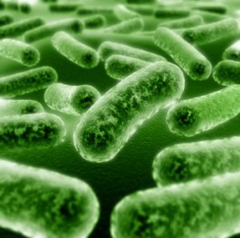
FLAGELLA
A large
number of bacteria possess flagella. Flagellum is the organ of locomotion. Flagellated bacteria are motile. Nonflagellated
bacteria are non-motile.
Examples:
Motile Bacteria: Escherichia, Salmonella, Proteus, Pseudomonas, Vibrio, Aeromonas, Plesiomonas, Campylobacter, Helicobacter, Clostridium tetani.
Nonmotile Bacteria: All Cocci, Shigella, Klebsiella, Corynebacterium diphtheriae, Clostridium perfringens, Bacillus anthraces, Haemophilus, Bordetella, Brucella, Yersinia, Pasteurella, Francisella.
Structure . Flagella are long, slender thread-like (12-30 nm in diameter) organs of locomotion. A flagellum is attached to the bacterial cell body by a complex structure consisting of filament, hook and basal body. The basal body has one pair of rings in Gram-positive bacteria and two pairs in Gram-negative bacteria. A bacterial flagellum is composed entirely of protein. A flagellum is made up of several thousand molecules of a protein subunit called flagellin. The flagellum is formed by the aggregation of subunits to form a helical structure.
A flagellated bacterium may become non-flagellated but not the reverse. The arrangement of flagella for a particular bacterium is constant and is one of the four types.
1. Peritrichous. Flagella are distributed over the entire cell, e.g. Escherichia coli, Salmonella genus.
2. Amphitrichous. There is a single flagellum at each pole.
3. Lophotrichous. There is a bunch of flagella at one or both poles, e.g. Spirillum minus.
4. Monotrichous. There is a single polar flagellum at one pole, e.g. Vibrio cholerae.
They have no function in the virulence of the bacteria. Antigenic structure of the flagella (H antigens) may be used for identification and classification of bacteria.
Sensory transduction is the behavior of the flagellated bacteria in response to the environment. This may be: (1) Chemotaxis is the movement of the bacterium towards a chemical attractant, e.g. sugar, amino-acid, (2) Aerotaxismovement towards the optimal oxygen concentration, (3) Electron acceptor taxis- movement towards alternative electron receptors, (4) Phototaxis- movement towards light of photosynthetic bacteria.
Motility Test. Hanging-drop preparation is done by placing a drop of bacterial suspension between a slide and cover slip separated by a circular layer of vaseline on the surface of the slide.
Flagella Staining. They are demonstrated by special staining. Flagella are not visible under light microscope. Bacteria are first treated with tannic acid salt which precipitates on flagella and cell walls and increases the diameter of the flagella. They are then visible under light microscope.
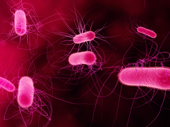
PILI (FIMBRIAE)
Pill or fimbriae are very thin, hair-like short rigid surface
appendages. Many Gram-negative bacteria possess pili or fimbriae. There
may be 100-500 pili in one cell. They are shorter and finer than
flagella and composed of protein subunits termed pilins. Minor proteins,
located at the tips of pili, are responsible for the attachment
properties. They are seen by electron microscope. Pellicle formation in
the fluid media by bacteria is due to fimbriae. Pill of different
bacteria are antigenically distinct. Some bacteria, e.g. N. gonorrhoeae are able to make pili of different antigenic types (antigenic variation) and
this can still adhere to cells by a type in presence of antibodies to
other types. Fimbriae of group A streptococci are the site of the
surface antigen, M protein. Lipoteichoic acid, associated with these
fimbriae is responsible for adherence of these streptococci to
epithelial cells of the hosts. There are two types of pili on function:
1. Ordinary pill. These play roles in adherence of bacteria to host cell surfaces. 'Colonization antigens' are ordinary pili that provide the cells with adherent properties, genetically determined by plasmids.
2. Sex pill. These give the 'maleness'. They are few in number and very long, e.g. in enterobacteria. During bacterial conjugation the male (donor) cell adhere with its sex pili and transfer DNA to non-male (recipient) cell. The pili act as conjugation tubes. In this way the genetic material which determines antibiotic resistance may pass from one bacterium to another. Sex pili possess receptors to which bacteriophage can become attached.
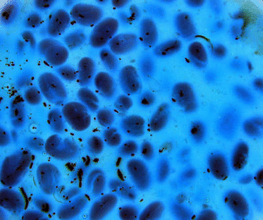
BACTERIAL SPORES
(ENDOSPORES)
Bacterial endospores are
formed when conditions for vegetative growth are not favourable.
Spore-Forming Bacteria
1.Bacillus and Clostridium genera form spores.
2. Sporosarcina (Gram-positive coccus) and possibly Coxiella burnetii can produce spores.
Demonstration of Spore:
1.In Gram stain, the spore appears as unstained colourless areas in cells.
2.Spores are stained with malachite green, or carbol fuchsin (by modified Ziehl-Neelsen stain).
Nature. The spore is a resting cell. Spores are in a subdued metabolic state and non-reproducing. Each cell forms a single internal spore that is liberated when the mother cell undergoes autolysis. The spore is highly resistant to heat, chemical agents and desiccation. In sporulation, each vegetative cell forms only one spore, and in subsequent germination each spore gives rise to a single vegetative cell.
