_SHAPE, SIZE AND ARRANGEMENT OF BACTERIA
Bacteria are classified into coccus and bacillus:
Coccus (Pleural- Cocci): A coccus is a round or oval shaped bacterium. In most cocci, one axis of the cell is almost equal to any other axis. But a coccus may be slightly oval, pointed at the ends; lancet, bean or kidney shaped (Gonococcus). Typical cocci are about 0.5-1.0 pm in diameter. During multiplication, cocci may arrange in irregular groups, chains, pairs or singles.
(a) Cocci in irregular groups
(b) Cocci in chains
(c) Cocci in pairs are called diplococcus.
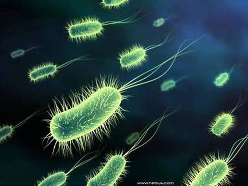
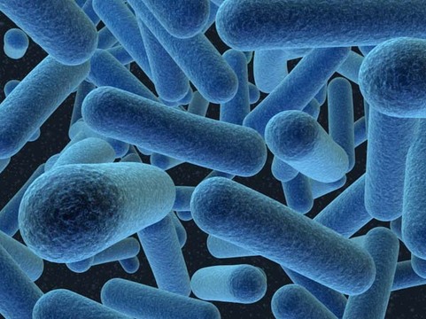
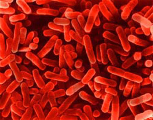
Bacillus (Pleural- Bacilli) or Rod. Bacillus is rod shaped. Bacterium having one axis of the cell longer than the other. Size : 1-5 Iim x 0.5-1.0 pm. Rods are mostly arranged singly. When the size of a rod is short and approximates to coccal form, it is called coccobacillus. Vibrio is comma-shaped single curved rod. Actinomyces and Nocardia are mycelial, filamentous or rods. Corynebacterium is club-shaped rod. Spirochaetes are long slender flexible spiral rods with several curves. Most are 5-20 pm x 0.1-0.2 gm. Fusobacterium is spindle shaped rod. Spirillum is spiral rod with several curves. Mycoplasma lack cell wall. Chlamydiae and Rickettsiae are strict intracellular bacteria.
On Gram-staining, cocci and bacilli (except Mycobacterium) are classified into Gram-positive bacteria and Gram-negative bacteria. This difference in the staining depends on the composition of bacterial cell wall. It is used to study the shape, size and arrangement of bacteria beside the staining character.
Pleomorphism indicates that there is considerable variation in the size and/or shape of a strain.
Involution form indicates that there is morphologically different forms of a strain. Most commonly occurs in the culture of Corynebacterium.
Pleomorphism indicates that there is considerable variation in the size and/or shape of a strain.
Involution form indicates that there is morphologically different forms of a strain. Most commonly occurs in the culture of Corynebacterium.
_CELL STRUCTURE OF BACTERIA
(Prokaryotic Cell Structure)
1. Cell Envelope: Layers that surround prokaryotic cell.
(a) Cytoplasmic or cell membrane. It is the innermost layer present in all bacteria which surrounds the cytoplasm.
(b) Cell wall. The layer between cytoplasmic membrane and capsule. It is present in all bacteria except mycoplasma, i.e. it is absent in mycoplasma.
(c) Capsule and Glycocalyx : Outermost layer. Some bacteria may have capsule and glycocalyx.
(d) S-layers.
2. Genetic Elements
(a) Nucleoid. Nucleoid is present in all bacteria.
(b) Plasmid. Plasmid is present in certain bacteria.
(c) Transposons are present in certain bacteria.
3. Cytoplasm. It is present in all bacteria.
4. Flagella. May or may not be present.
5. Pili (Fimbriae). May or may not be present.
6. Spores. Endospores are formed by certain bacteria.
CELL ENVELOPE
The layers that surround the bacterial cells are referred collectively as the cell envelope. Cell envelope consists of cytoplasmic membrane, cell wall, and for some bacteria capsule and glycocalyx from inner to outer surface. Many bacteria may have an S-layer as the outermost component of the cell envelope. Cell envelope differs in Gram-positive and Gram-negative bacteria as follows:
1. Gram-Positive Cell Envelope. Cell envelope consists of two to three layers: (a) Cytoplasmic membrane, (b) Cell wall-thick peptidoglycan layer: May be 40 sheets, and (c) Capsule or an S-layer in some bacteria.
2. Gram-Negative Cell Envelope. Highly complex, multilayered envelope: (a) Cytoplasmic membrane or inner membrane, (b) Cell wall- peptidoglycan- only one or two sheets, (c) Outer membrane, (d) Capsule or S-layer may also be present, (e) Periplasmic space. The space between the inner and outer membrane is called periplasmic space.
_Cytoplasmic Membrane
Structure : Cytoplasmic membrane is the innermost layer of cell envelope that externally limits cytoplasm. It is composed of phospholipids and proteins.
Mesosomes are convoluted invaginations of the cytoplasmic membrane and visible with electron microscope. There are two types: septa) mesosomes and lateral mesosomes. Septa) mesosome function in the formation of cross wall during cell division.
Functions : (1) Selective permeability and transport of solutes, (2) bearing the receptors and other proteins of the chemotactic system, (3) bearing the enzymes and carrier molecules that function in the biosynthesis of DNA, membrane lipids, and cell wall polymers, (4) electron transport and oxidative phosphorylation in aerobic species, and (5) secretion of hydrolytic enzymes and toxins.
Bacterial chromosome (DNA) is attached to a septa) mesosome during cell division.
Cell Wall
Layers of the cell envelope lying between the cytoplasmic membrane and capsule is the 'cell wall'. It is external to cytoplasmic membrane. A bacterial cell wall is prokaryotic. Mycoplasmas have no cell wall. Functions: The cell wall is mostly rigid but also exhibit tensile strength. The strength depends on peptidoglycan, murein or mucopeptide which is the main component. The cell wall maintains the bacterial shape and protects the cell from lysis. The cell wall plays an important part in cell division. During cell division, a transverse partition is formed from the cell wall by its inward growth. Many antibacterial agents inhibit cell wall synthesis. Differential Gram-staining character leading to classification into Gram- positive and Gram-negative bacteria reside in the cell-wall. Peptidoglycan layer is common component, others are special components.
1. Cell-walls of Gram- positive bacteria
(a) Peptidoglycan Layer. Peptidoglycan comprise up to 50% of the cell wall material and may be 40 sheets. It is composed of N-acetyl muramic acid, N-acetyl glucosamine and peptides. Lysozyme acts on it,
(b) Teichoic acids. Teichoic acids lie on the outside surface of the peptidoglycan layer in most Gram positive bacteria. Teichoic acids are polymers containing ribitol or glycerol. There are two types of teichoic acid-wall teichoic acid linked to peptidoglycan and membrane teichoic acid linked to membrane.
(c) Polysaccharides.
2. Cell walls of Gram-negative bacteria:
(a) Peptidoglycan Layer. Peptidoglycan comprises 5 to 10% of the wall material. Only one or two sheets.
(b) Lipoprotein. Lipoprotein is the most abundant protein of Gram negative cells. It probably stabilizes the outer membrane.
(c) Outer membrane. Lipid in nature. Serves to protect the cell of enteric bacteria from bile salts.
(d) Lipopolysaccharide (LPS). LPS molecule is toxic and is referred to as endotoxin of Gram negative bacteria. LPS is split into lipid A and polysaccharide. Lipid A is the toxin, and polysaccharide is the major surface antigen of the bacterial cell- called 0 antigen. Lipooligosaccharides (LOS) are important virulence factors and are antigenic.
(e) Periplasmic space. The space between the inner and outer membrane. It contains gel-like solutions of proteins and murein layer. Periplasmic proteins include (a) binding proteins for specific substrates like vitamins, sugar, amino acids, ions, and (b) enzymes like P-lactamase, alkaline phosphatase.
Capsule And Glycocalyx
Many bacteria synthesize extracellular polymers forming variable outer layer when growing in their natural environment. (i) Capsule. Capsule forms a well-defined layer closely surrounding the cell. Functions: Capsule plays role in the virulence of pathogenic bacteria by contributing to invasiveness and preventing phagocytosis. Typing of certain bacteria are done on antigenic character of the capsule. Capsulated bacteria form smooth or mucoid colonies, and if they become noncapsulated then form rough colonies, e.g. Streptococcus pneumoniae, Klebsiella pneumoniae. The extracellular material is polysaccharide except the capsule of Bacillus anthraces which is poly D-glutamic acid, a polypeptide. Capsule is demonstrated by special stain. (2) Glycocalyx. When the polymer forms a loose meshwork of fibrils, it is called glycocalyx. Glycocalyx plays role in adherence of bacteria on surface, e.g. Streptococcus mutans adheres to tooth enamel. (3) If the polymer is detached from the cells then it is called "slime layer". Bacteria may be trapped in slime layer.
Capsule Stain. The bacterial film on the slide is stained with hot crystal violet solution followed by rinsing with copper
sulphate solution. The cell and background appear dark blue and the capsule a much paler blue.
Examples:
Capsulated Bacteria: Streptococcus pneumoniae, Kleb-iella pneumoniae (large capsule), Bacillus anthraces. Staphylococcus (some strains), Streptococcus pyogenes (some strains), Bordetella pertussis.
Non-capsulated Bacteria: Neisseria, Corynebacterium, Salmonella, Shigella, Vibrio, Proteus, Pseudomonas.


